






Exosomes are often described as “messengers” between cells and one of the key directions in regenerative aesthetics. But the word exosomes on a serum label or in a treatment protocol can refer to very different materials. Here is what human studies have already shown, where the evidence ends, and where marketing begins.
Exosomes are a subtype of extracellular vesicles that cells use to transmit biological signals. In small clinical studies, certain exosome-based products improved skin hydration, elasticity, texture, and recovery, including after laser procedures and microneedling.
The interest in exosomes did not come out of nowhere. Cells do release membrane-bound particles into their surroundings, and these particles can carry proteins, lipids, and nucleic acids. Through them, cells exchange signals involved in inflammation, healing, and tissue remodeling. This is why extracellular vesicles are being studied in regenerative medicine, and why aesthetic medicine is trying to use this mechanism to improve skin quality.
Exosomes entered cosmetology from cell biology, but on the market the term is used much more broadly. The word exosomes on a label may refer to products that differ in origin and composition, so they should be assessed by the specific formula and the way they are used. In regenerative cosmetology and skin repair, this is essential: a serum, a product applied after a procedure, and an injectable product are different scenarios with different levels of evidence and risk.
What exosomes are — and why the name is not always accurate
Extracellular vesicles are small membrane-bound particles that cells release into their environment. They can form in different ways. Exosomes first form inside the cell: future vesicles accumulate in multivesicular bodies, which then fuse with the cell membrane and release them outside. Another type of extracellular vesicle forms directly on the cell surface: a section of the plasma membrane buds outward and separates together with part of its contents.
Once released from the cell, particles of different origins may look similar in size and appearance. This means it is not always possible to determine, from the finished vesicle alone, that it was formed specifically inside the endosomal system and is therefore an exosome. For this reason, the international MISEV2023 guidelines recommend using the broader term “extracellular vesicles” when their origin has not been established. For the cosmetics market, this has practical importance: the word exosomes on a label does not yet prove that the product actually contains exosomes.
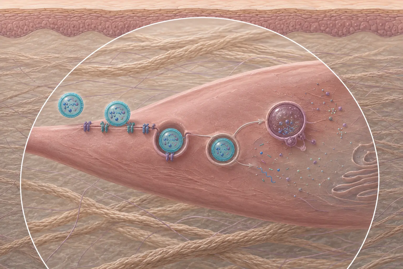
Fig. Schematic illustration of exosome interaction with skin cells
What may actually be inside an “exosome” product
In studies and finished products, the word “exosomes” may refer to different biological materials. These may be isolated and purified extracellular vesicles, a cell secretome, or conditioned medium containing a whole complex of substances released by cells. Vesicles and vesicle-like nanoparticles of plant origin form a separate category. These terms are not interchangeable, and the composition of such materials can differ significantly.
Extracellular vesicles (EVs) are isolated from various biological sources. Clinical and laboratory studies examine vesicles from mesenchymal stromal cells, platelets, microorganisms, milk, and other materials. Their properties depend on the source, the condition of the cells, culture conditions, and the methods used for isolation, purification, and storage.
The secretome is a much broader concept. It refers to the total set of substances and particles that cells release: proteins, growth factors, cytokines, metabolites, and extracellular vesicles. Exosomes, if their origin is confirmed, are only one component of the secretome.
Conditioned medium is not the name of the total set of components released by a cell, but the material obtained during cell culture. Cells spend a certain amount of time in a nutrient medium and release components of their secretome into it. The medium is then collected and, depending on the technology, purified, concentrated, or used for further isolation of individual fractions. This is why the effect of conditioned medium cannot be explained by exosomes alone: along with vesicles, it may contain other biologically active components.
A separate story is the so-called “plant exosomes”. Plants also have extracellular vesicles, and vesicle-like nanoparticles can be obtained from plant materials. If the formation mechanism of specific particles has not been established, calling them exosomes is incorrect. It is more accurate to speak of plant extracellular vesicles or vesicle-like nanoparticles. They may be a research subject in their own right, but they are not a plant analogue of human exosomes with automatically identical properties.
How extracellular vesicles may affect the skin
The idea behind exosome therapy is not that vesicles turn into new cells. They are not stem cells and cannot replace damaged tissue. They are viewed as carriers of biological signals. After contact with a target cell, a vesicle may interact with its membrane or be taken up by the cell, while the molecules it carries may alter the activity of signaling pathways.
In cell cultures and wound-healing models, extracellular vesicles from different sources have affected the activity of keratinocytes and fibroblasts, inflammatory signals, angiogenesis, and components of the extracellular matrix. This is where claims about “collagen stimulation,” “accelerated regeneration,” and “cellular rejuvenation” come from.
The composition of vesicles is not universal either. Even EVs from the same type of cells, obtained and purified using different protocols, can differ. This means “exosomes” cannot be assessed as one standardized active ingredient in the way a specific retinoid molecule can.
What human studies have already shown
A 2026 systematic review included 19 clinical studies of exosome-based and EV protocols for skin rejuvenation. They described short-term improvements in hydration, elasticity, wrinkles, pores, pigmentation, and overall skin appearance. This is already more than a purely laboratory hypothesis. At the same time, most of the included studies were not randomized, the products and methods of use differed, and follow-up was often short. Because of this heterogeneity, the authors did not perform a meta-analysis.
There are also controlled studies. In a 2023 randomized split-face study, 28 participants underwent three microneedling sessions. After the procedure, one side of the face received a solution containing vesicles obtained from conditioned medium of adipose-derived mesenchymal stromal cells, while the other side received saline. After 12 weeks, the side treated with the study product received better scores on the Global Aesthetic Improvement Scale (GAIS). This points to an additional effect of the studied formula when combined with microneedling.
Another illustrative example is a randomized, double-blind split-face study of 25 people with atrophic post-acne scars. After three sessions of fractional CO2 laser, one side was treated with a gel containing extracellular vesicles derived from adipose stromal cells, while the other side received a control gel. After 12 weeks, the ECCA scar severity score decreased by 32.5% versus 19.9% on the control side; erythema was also less pronounced, and recovery was shorter. In other words, when the exosome-based product was combined with laser treatment, tissue regeneration in the scar area was better.
So the evidence base has already moved beyond laboratory experiments, but it is still early. There is not yet enough data to establish the optimal EV source, a standard dose, a universal number of procedures, or a reliable duration of effect.
Serum, microneedling, and injection: three different scenarios
The result depends not only on what the product contains, but also on where it goes. The stratum corneum performs its main function well: it limits the penetration of large and hydrophilic structures. So the mere presence of extracellular vesicles in a cream or serum does not prove that functional particles will reach the living layers of the skin in sufficient quantities. A topical product may still improve hydration, comfort, and barrier function thanks to the formula as a whole.
Why exosomes are often combined with laser or microneedling
After fractional laser or microneedling, the barrier is temporarily disrupted, and the pathway for substances entering the skin changes. That is why many positive clinical studies examine EVs specifically as an add-on to a procedure. However, it is important to remember that both laser and microneedling trigger remodeling on their own, and in uncontrolled studies it is difficult to separate the contribution of vesicles from the effect of the procedure itself. In split-face studies, the side treated with the control product makes it possible to assess the additional effect of the studied formula, but not the action of extracellular vesicles separately from microneedling or laser.
A disrupted barrier also changes the safety requirements. A product applied over open microchannels must be specifically intended by the manufacturer for this type of use and must meet requirements for quality, purity, and microbiological safety. A home-use serum is not suitable for delivery through microchannels, even if the packaging says exosomes. Only products specifically developed for this purpose should be used.
Exosome injections: direct delivery into the dermis and its risks
During an intradermal injection, the product enters the dermis directly — into the extracellular space next to fibroblasts, endothelial cells, and immune cells. Extracellular vesicles may bind to receptors on the surface of these cells or be taken up by them through endocytosis. The proteins, lipids, and RNA contained in vesicles can influence signaling pathways related to inflammation, cell migration, and remodeling of the extracellular matrix. The outcome depends on the vesicle source, their molecular cargo, the dose, and the purity of the specific product.
The argument in favor of injections is the ability to deliver a defined volume of product directly to living tissue without relying on penetration through the stratum corneum. This sets the method apart from a conventional serum and from application after laser or microneedling. In a small clinical study, 25 participants received a single injection of their own extracellular vesicles obtained from blood. After five days and three weeks, the researchers recorded improvements in hydration, elasticity, texture, and several other skin parameters without any reported adverse reactions. At the same time, the study had no control group and follow-up lasted only three weeks, so it shows the promise of the approach but does not yet establish its long-term effectiveness.
The downside of direct injection is that the entire formula enters the dermis along with the vesicles: proteins, stabilizers, preservatives, residual cellular components, and possible impurities. If the body recognizes one of the components as foreign or cannot break it down quickly, prolonged inflammation with papules, nodules, or granulomas may develop. On top of this are the usual risks of any injection procedure: pain, swelling, bruising, infection, and vascular injury.
Such complications have already been described in the clinical literature. In 2024, skin necrosis was reported after intradermal injection of a lyophilized exosome product. In a 2025 case series, four female patients developed persistent redness, nodules, granulomatous inflammation, and scarring after injection of unregulated exosome formulations.
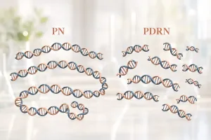
In terms of the delivery method, such a procedure may resemble other skin boosters, but it should not be equated with PN and PDRN injections. PN, PDRN, and extracellular vesicles differ in structure, mechanisms of action, manufacturing requirements, and evidence base.
Regulatory status is also determined not by the word exosomes on the packaging, but by the product composition and its approved method of use. The FDA states that there are no approved exosome products in the United States. In the EU, injectable administration is not considered cosmetic use, and cells, tissues, and products of human origin are included in the list of prohibited cosmetic ingredients. So the key question before a procedure is whether the specific product is registered for injection and what exactly is inside the vial.
What to check in an exosome product
The claim “5 billion exosomes” may sound convincing, but the number itself says little about product quality. The result depends on the counting method, product purity, and the proportion of non-vesicular particles; the number also does not show how many vesicles retain biological activity.
For a professional product, it is far more useful to know:
- what source the material was obtained from;
- whether it is purified extracellular vesicles, a secretome, conditioned medium, or another raw material;
- how the manufacturer characterizes particle size, quantity, markers, and purity;
- how sterility, stability, and storage conditions are confirmed;
- which method of use the product is officially intended for;
- whether there is a clinical study of the finished formula itself, not only cell experiments;
- what the result was compared with: placebo, the procedure without the product, or only the skin condition before treatment;
- how many people participated and how long the result was followed.
For a home-use product, the criteria are simpler. Hydration, reduced dryness, or improved barrier function may be real results of using exosome-based cosmetics in cream, serum, essence, and similar formats.
Extracellular vesicles are a real field of biology and regenerative medicine, and clinical studies are already showing positive signals for recovery after procedures and for certain skin quality parameters. However, between EV biology and a specific ampoule on a cosmetologist’s tray lie manufacturing, purification, particle characterization, storage, method of use, and clinical validation. These are the data by which a product should be assessed — not by the word exosomes on the label.
Sources
- Welsh J. A., Goberdhan D. C. I., O'Driscoll L. et al. Minimal information for studies of extracellular vesicles (MISEV2023): From basic to advanced approaches. Journal of Extracellular Vesicles, 2024; 13: e12404.
- Flores Rodríguez J. C., Toledo Avelar L. E., Yi K. et al. Efficacy of Exosome-Based Therapies for Skin Rejuvenation: A Systematic Review of Human Studies. Cureus, 2026; 18(2): e104182.
- Park G. H., Kwon H. H., Seok J. et al. Efficacy of combined treatment with human adipose tissue stem cell-derived exosome-containing solution and microneedling for facial skin aging: A 12-week prospective, randomized, split-face study. Journal of Cosmetic Dermatology, 2023; 22(12): 3418-3426.
- Kwon H. H., Yang S. H., Lee J. et al. Combination Treatment with Human Adipose Tissue Stem Cell-derived Exosomes and Fractional CO2 Laser for Acne Scars: A 12-week Prospective, Double-Blind, Randomized, Split-Face Study. Acta Dermato-Venereologica, 2020; 100: adv00310.
- Park K. Y. Adverse Reactions Following Intradermal Injection of Exosome-Based Formulations: A Case Series. Journal of Cosmetic Dermatology, 2025; 24(10): e70520.
- Tawanwongsri W., Vachiramon V. Skin necrosis after intradermal injection of lyophilized exosome: A case report and a review of the literature. Journal of Cosmetic Dermatology, 2024; 23(5): 1597-1603.
- U.S. Food and Drug Administration. Consumer Alert on Regenerative Medicine Products Including Stem Cells and Exosomes. 2020.
- Regulation (EC) No 1223/2009 of the European Parliament and of the Council on cosmetic products. Annex II.